Internal decapitation, a term that invokes fear and curiosity, is a rare yet severe medical condition that requires immediate attention. Despite its alarming name, it does not involve an actual separation of the head from the body, but rather a significant injury to the ligaments that connect the skull to the spine. This injury, often resulting from traumatic events like car accidents, sports injuries, or falls, can have devastating consequences if not promptly diagnosed and treated. In this article, we'll delve deep into the anatomy, causes, diagnosis, treatment, and recovery associated with internal decapitation, aiming to provide a thorough understanding of this critical condition.
Internal decapitation, medically known as atlanto-occipital dislocation, occurs when the ligaments connecting the skull and the atlas (the first cervical vertebra) are severely damaged, causing instability. This instability can lead to a range of complications, including spinal cord injury, paralysis, or even death. The complexity of this injury lies in its subtle nature; often, the symptoms are not immediately evident, making timely diagnosis a challenge. Advanced imaging techniques, such as MRI and CT scans, play a crucial role in identifying the extent of the injury and guiding treatment decisions.
The journey to recovery from internal decapitation is often long and arduous, requiring a multidisciplinary approach involving neurologists, orthopedic surgeons, and rehabilitation specialists. Treatment typically involves surgical stabilization of the cervical spine, followed by intensive rehabilitation therapy to regain strength and mobility. While the prognosis varies depending on the severity of the injury and the promptness of medical intervention, many patients can achieve significant recovery with appropriate care. This article aims to shed light on the intricacies of internal decapitation, offering valuable insights into its management and emphasizing the importance of awareness and preparedness in preventing such life-threatening injuries.
Table of Contents
- Anatomy of the Cervical Spine
- What is Internal Decapitation?
- Causes of Internal Decapitation
- Symptoms and Signs
- Diagnosing Internal Decapitation
- Treatment Options
- Surgical Interventions
- Rehabilitation and Recovery
- Prognosis and Outcomes
- Preventing Internal Decapitation
- Impact on Quality of Life
- The Role of Research and Innovation
- Real-Life Case Studies
- Support and Resources
- Frequently Asked Questions
Anatomy of the Cervical Spine
The cervical spine, a marvel of biological engineering, is composed of seven vertebrae known as C1 to C7. It plays a crucial role in supporting the skull, facilitating head movement, and protecting the spinal cord. The first cervical vertebra, the atlas, and the second, the axis, are unique compared to other vertebrae. They form the atlanto-occipital joint, allowing for the nodding motion of the head, and the atlanto-axial joint, enabling head rotation.
The cervical spine's structure is designed to be both strong and flexible, supported by ligaments that provide stability and allow for a range of motion. These ligaments include the alar and transverse ligaments, among others, which are vital in preventing excessive movement that could damage the spinal cord. Understanding the anatomy of the cervical spine is essential for grasping the implications of internal decapitation.
Internal decapitation involves the disruption of these critical ligaments, which can lead to instability at the cranio-cervical junction. The delicate nature of the structures involved means that even slight displacements can have severe consequences, including neurological deficits or fatal outcomes if the injury compromises the spinal cord.
What is Internal Decapitation?
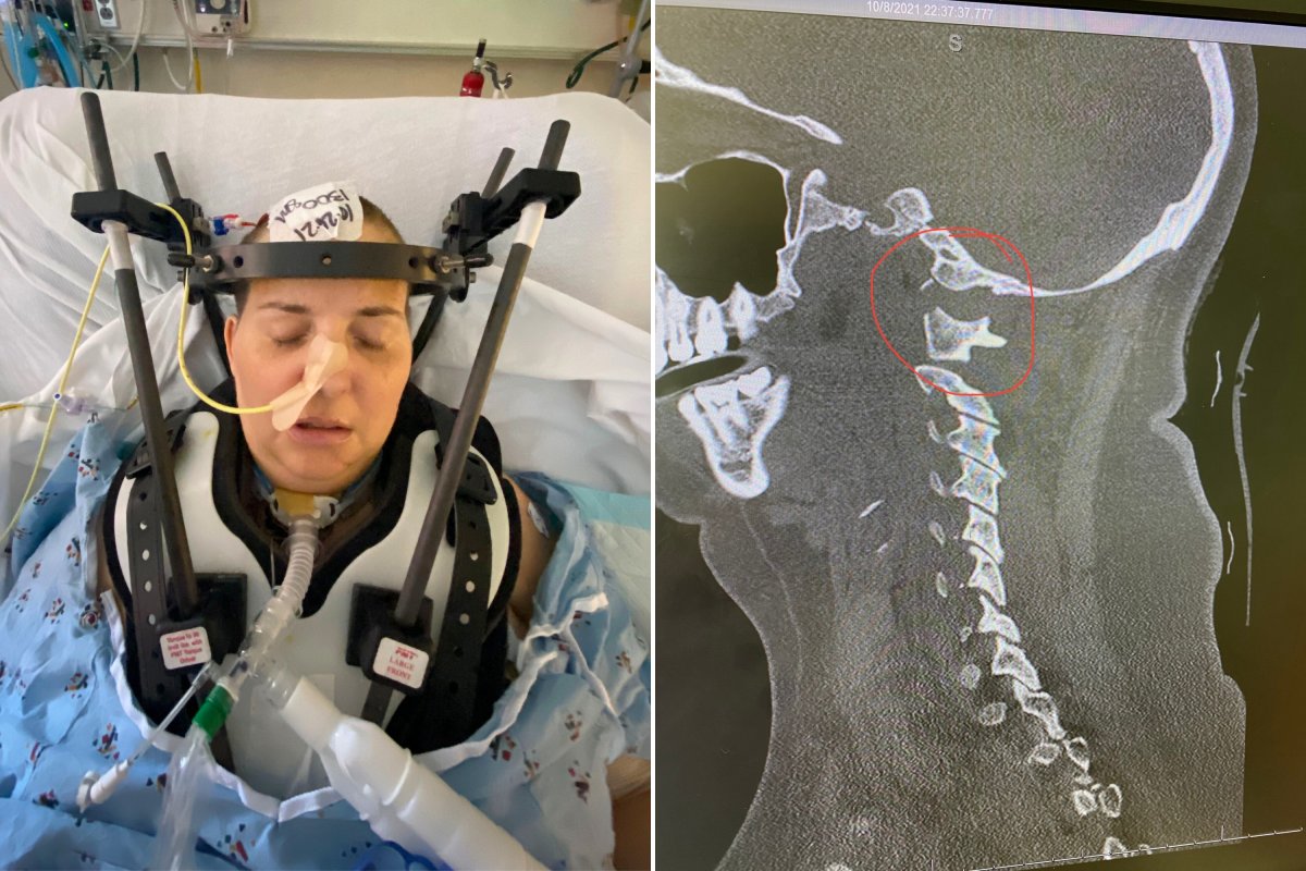
Internal decapitation, also known as atlanto-occipital dislocation, is a traumatic injury where the ligaments between the skull and the cervical spine are severely compromised. Unlike an external decapitation, where the head is entirely severed, internal decapitation involves an internal disconnection that can be just as life-threatening.
The term 'internal decapitation' highlights the severity of the injury, as it often requires urgent medical intervention to stabilize the patient and prevent permanent damage. The condition is rare, but when it occurs, it is predominantly seen in high-impact trauma cases, such as vehicle collisions, falls from significant heights, or during contact sports.
Despite its rarity, understanding internal decapitation is crucial due to its potentially fatal nature. Prompt recognition and treatment can be the difference between life and death, emphasizing the need for awareness among both medical professionals and the general public.
Causes of Internal Decapitation
Internal decapitation is primarily caused by traumatic events that exert extreme forces on the neck, leading to the disruption of the ligamentous connections between the skull and spine. The most common causes include motor vehicle accidents, where sudden acceleration or deceleration forces impact the head and neck.
Sports injuries, particularly in contact sports like football, rugby, or wrestling, can also lead to internal decapitation. These activities often involve high-speed collisions or falls that can compromise the cervical spine's stability. Additionally, falls from heights, especially when landing on the head or neck, are significant risk factors.
While these are the primary causes, internal decapitation can also occur in cases of severe physical assault or during certain medical procedures, albeit rarely. Understanding these causes is essential for prevention and prompt diagnosis, particularly in high-risk scenarios.
Symptoms and Signs
Identifying internal decapitation can be challenging due to its subtle presentation and the overlap of symptoms with other neck injuries. Common signs include severe neck pain, loss of consciousness, respiratory difficulties, and neurological deficits such as paralysis or altered sensation.
In some cases, symptoms may be delayed, making diagnosis difficult. Patients might experience dizziness, difficulty swallowing, or changes in voice. Due to the potential for severe complications, any suspicion of internal decapitation warrants immediate medical evaluation.
Advanced imaging techniques like CT scans or MRI are essential for diagnosing internal decapitation. These tools provide detailed views of the cervical spine, allowing for accurate assessment of ligamentous injuries and the extent of displacement.
Diagnosing Internal Decapitation
Diagnosing internal decapitation requires a high index of suspicion, particularly in trauma cases. The primary diagnostic tools include radiographic imaging, with CT scans and MRIs providing the most detailed views of the cranio-cervical junction.
CT scans are often the first choice in emergency settings due to their speed and ability to detect bony injuries and gross ligamentous disruptions. MRI, on the other hand, offers superior visualization of soft tissues, including ligaments, and is crucial for confirming the diagnosis and planning surgical intervention.
In addition to imaging, clinical assessment of neurological function and stability is vital. This approach ensures a comprehensive evaluation, enabling healthcare professionals to determine the most appropriate treatment strategy.
Treatment Options
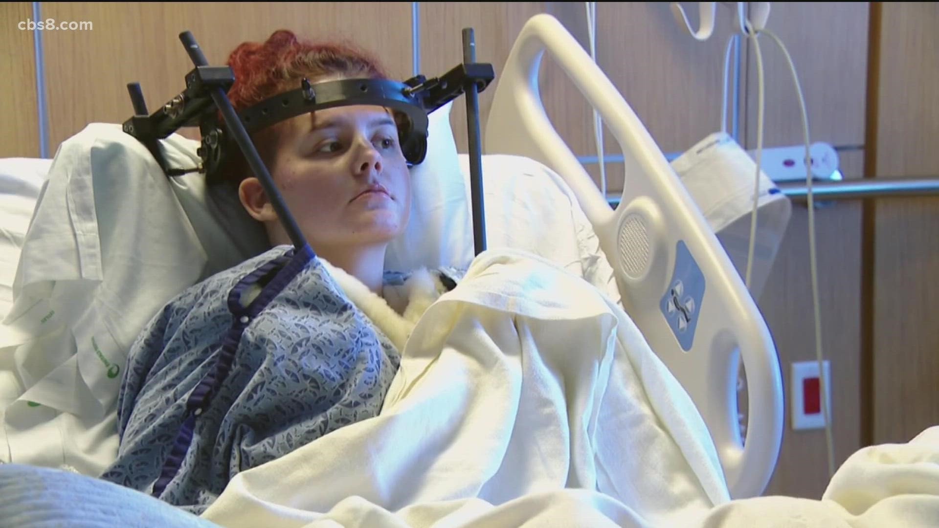
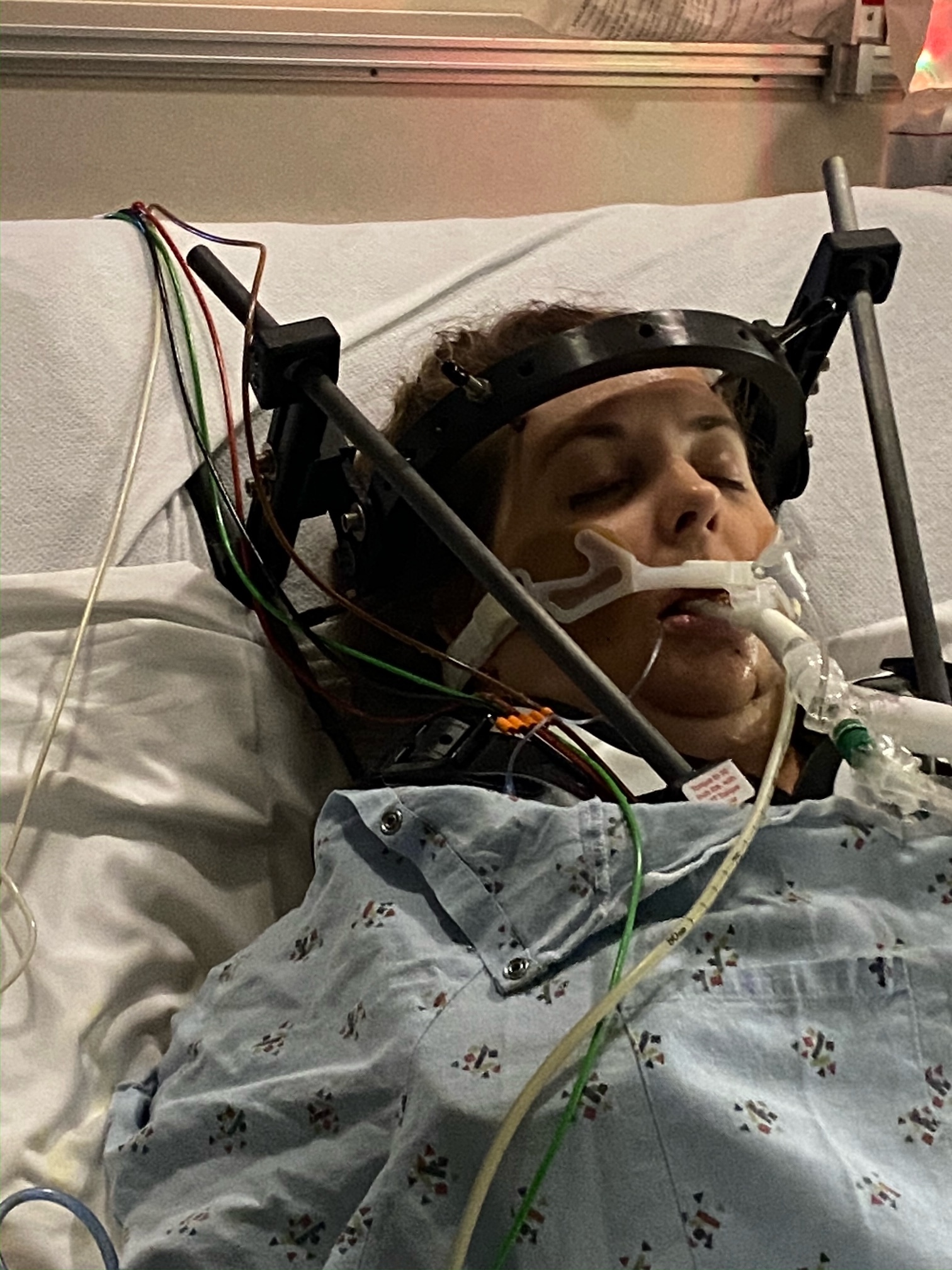
Treatment of internal decapitation primarily involves stabilizing the cranio-cervical junction to prevent further injury and allow for healing. This often requires surgical intervention, particularly in cases with significant instability or neurological deficits.
Non-surgical management may be considered in less severe cases, involving immobilization using cervical collars or traction devices. However, due to the potential for catastrophic outcomes, surgical stabilization is frequently preferred to ensure long-term success and prevent recurrent instability.
Rehabilitation is a critical component of treatment, focusing on restoring function and mobility while minimizing pain. A multidisciplinary approach involving physical therapists, occupational therapists, and medical specialists is often necessary to achieve optimal recovery outcomes.
Surgical Interventions
Surgical intervention for internal decapitation typically involves cranio-cervical stabilization using various techniques, such as occipital-cervical fusion or the use of internal fixation devices. These procedures aim to realign and stabilize the skull and cervical spine, preventing further displacement and protecting the spinal cord.
The choice of surgical technique depends on the injury's severity and the patient's overall health. Surgeons must consider factors like the extent of ligamentous damage, the presence of other injuries, and the patient's ability to tolerate surgery.
Post-surgical care is crucial for successful recovery, involving close monitoring for complications like infection, hardware failure, or neurological deterioration. Rehabilitation is initiated as soon as medically feasible, focusing on regaining strength, mobility, and independence.
Rehabilitation and Recovery
Rehabilitation following internal decapitation is a comprehensive process aimed at maximizing functional recovery and improving the patient's quality of life. It typically involves a tailored program of physical therapy, occupational therapy, and, in some cases, speech therapy, depending on the extent of neurological involvement.
Physical therapy focuses on strengthening the neck muscles, improving range of motion, and enhancing balance and coordination. Occupational therapy aids in regaining independence in daily activities, while speech therapy may be necessary if there are difficulties with swallowing or communication.
Recovery timelines vary significantly based on the injury's severity and the individual's response to treatment. Some patients may achieve near-complete recovery within months, while others may require ongoing therapy and support to manage long-term deficits.
Prognosis and Outcomes
The prognosis for internal decapitation depends on various factors, including the injury's severity, the speed of intervention, and the presence of associated injuries. Early diagnosis and treatment are critical for improving outcomes and minimizing the risk of permanent disability or death.
Many patients experience significant recovery with appropriate medical and rehabilitative care, although some may face long-term challenges such as chronic pain, limited mobility, or neurological deficits. Ongoing support and therapy can help manage these issues and enhance quality of life.
Advances in medical technology and surgical techniques continue to improve outcomes for those with internal decapitation, offering hope for recovery even in severe cases. Research and innovation remain vital in enhancing treatment options and promoting better prognostic outcomes.
Preventing Internal Decapitation
Prevention of internal decapitation focuses on minimizing the risk of traumatic events that could lead to such injuries. This involves promoting safety measures in high-risk activities, such as wearing seat belts in vehicles, using protective gear in sports, and employing fall prevention strategies in the workplace and at home.
Education and awareness are essential components of prevention, emphasizing the importance of recognizing and responding to potential neck injuries promptly. Training programs for medical professionals and first responders can enhance their ability to identify and manage internal decapitation cases effectively.
Incorporating safety technologies in vehicles, such as advanced restraint systems and collision avoidance features, can also play a significant role in preventing internal decapitation and other severe injuries.
Impact on Quality of Life
Internal decapitation can have a profound impact on a patient's quality of life, affecting physical, emotional, and social well-being. The injury may lead to chronic pain, reduced mobility, and challenges in performing everyday activities, significantly affecting independence and self-esteem.
Psychosocial support is crucial for patients and their families, helping them cope with the emotional and mental challenges associated with the injury. Support groups, counseling, and rehabilitation programs can provide valuable resources and assistance in navigating life after internal decapitation.
Despite the challenges, many patients show remarkable resilience and adaptability, achieving meaningful recovery and regaining a sense of normalcy with appropriate care and support.
The Role of Research and Innovation
Research and innovation play a pivotal role in advancing the understanding and management of internal decapitation. Ongoing studies aim to improve diagnostic techniques, enhance surgical interventions, and develop novel rehabilitation strategies to optimize recovery outcomes.
Technological advancements, such as improved imaging modalities and minimally invasive surgical techniques, continue to refine treatment approaches, reducing risks and improving patient outcomes. Collaborative efforts among researchers, clinicians, and industry leaders are essential in driving progress and translating scientific discoveries into clinical practice.
The future of internal decapitation management holds promise, with continued research efforts paving the way for more effective prevention, diagnosis, and treatment strategies.
Real-Life Case Studies
Exploring real-life case studies of internal decapitation provides valuable insights into the challenges and triumphs faced by patients and healthcare providers. These stories highlight the importance of timely intervention, the effectiveness of treatment strategies, and the resilience of individuals in overcoming adversity.
Case studies can also shed light on the diverse presentations of internal decapitation, emphasizing the need for personalized treatment plans and multidisciplinary collaboration in achieving successful outcomes.
By sharing these experiences, we can foster a deeper understanding of internal decapitation and inspire hope and determination for those affected by this condition.
Support and Resources
For individuals and families affected by internal decapitation, access to support and resources is critical in navigating the recovery journey. Organizations and support groups offer valuable assistance, providing information, counseling, and a sense of community for those facing similar challenges.
Healthcare professionals play a key role in connecting patients with appropriate resources and support services, ensuring they receive comprehensive care and guidance throughout their recovery.
Empowering patients with knowledge and support can significantly enhance their ability to manage the physical and emotional aspects of internal decapitation, promoting resilience and improving quality of life.
Frequently Asked Questions
- What is the difference between internal and external decapitation? Internal decapitation involves a disconnection within the body, specifically at the cranio-cervical junction, whereas external decapitation is an actual separation of the head from the body.
- Can internal decapitation be prevented? While not all cases can be prevented, safety measures such as wearing seat belts, using protective sports gear, and practicing fall prevention strategies can reduce the risk.
- How is internal decapitation diagnosed? Diagnosis typically involves advanced imaging techniques like CT scans and MRIs, which provide detailed views of the cervical spine and adjacent structures.
- What are the long-term effects of internal decapitation? Long-term effects vary depending on the injury's severity but may include chronic pain, limited mobility, and neurological deficits. Ongoing rehabilitation and support can help manage these issues.
- Is surgery always required for internal decapitation? Surgery is often necessary to stabilize the cranio-cervical junction, although non-surgical management may be considered in less severe cases.
- What role does rehabilitation play in recovery? Rehabilitation is crucial for restoring function and mobility, involving physical therapy, occupational therapy, and other modalities to optimize recovery outcomes.
In conclusion, internal decapitation is a life-threatening condition that requires immediate medical attention and a comprehensive approach to treatment and recovery. Through education, research, and collaboration, we can enhance our understanding and management of this critical injury, improving outcomes and quality of life for those affected.
For further information and resources, please visit: Mayo Clinic - Spinal Cord Injury.